


A vascular assessment is necessary to determine the source of the varicose veins or spider veins in order to propose the best treatment.
Color echography with ultrasound measures the size of the vessels, the direction of the blood flow and allows the physician to determine the origin of the problem and to correlate it with the symptoms (heaviness - pain - cramps - itching…) or with the cutaneous signs (skin pigmentation - cyanosis - edema), which may also be produced by other diseases ( kidney, heart, thyroid, sciatic nerve ) .
There are 3 types of varicose veins:
1 - Telangiectasia ( spider veins ) can be isolated and treated promptly by micro-sclerotherapy.They produce a visual discomfort but no symptoms, only if independent of deeper dilated venous axis and producing a local or global congestion. They are common at thigh level and towards the knee. Cosmetics treatment cannot be claimed to your health insurance.
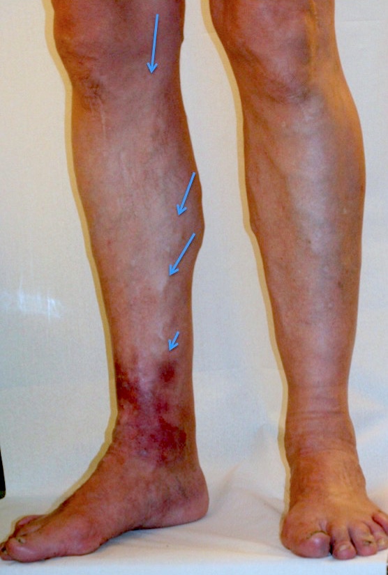
When spider veins accumulate in some peripheral area ( foot) , they are the sign of a venous hypertension or congestion which must be taken seriously. This build-up of venous pressure in deep tissue can result in slow skin healing after an injury , in bleeding or even in the build up of leg ulcers. It is usually related to the dysfunction of deep or superficial venous axes (saphenous veins) and may be associated to delays or obstructions in the arterial network. An ultrasound examination is mandatory before any action is taken.
The building up of a column of pressure creates a congestion (SEE HERE) of the venous network responsible for most symptoms and signs. A logical treatment is to get rid off the source and of the structures involved including perforator veins ( connecting the superficial system to the deep system) .
These techniques have been used for 15 years already. Medical insurances cover the procedure when performed by a USSMV ( Union Suisse des Sociétés Maladies Vasculaires ) certified specialist , see http://www.uvs.ch
The main axis can be removed by classic stripping, or welded by thermic devices using endovenous
LASER or RADIOFREQUENCY ( SEE video ) when hemodynamic criteriae allow it .
2 - Reticular varicose veins or branches are often dependent upon larger caliber venous axes such as saphenous veins. If their caliber remains moderate, and if the trunk that drains them is not too dilated, and remains local, a local phlebectomy crochet (hook?) is performed under local anesthetics. It involves the removal of small venous segments by making 1 mm openings every 4-5 cm, (e.g,knee and ) .
If varicose veins are smaller,with a diameter of 3 mm or less , a simple or foam sclerotherapy may be enough. Because the basic problem can evolve, main axis monitoring remains necessary in the long run